Vol. 8, No. 1
Jan. 2016

Photo feature – A year in review

The field of laboratory science saw many changes and achievements in 2015. Here’s a look at a few of these by Iowa’s environmental and public health laboratory.

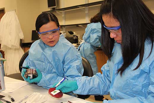
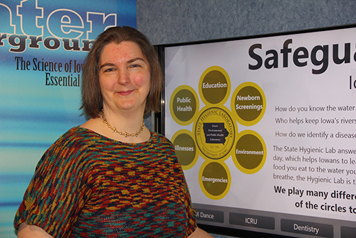
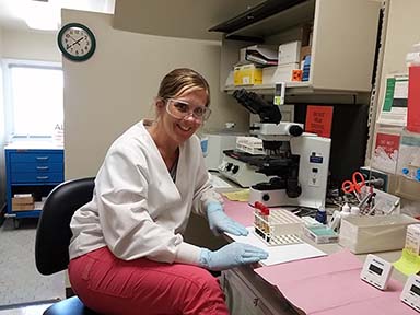
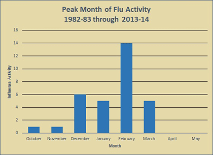
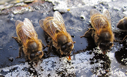
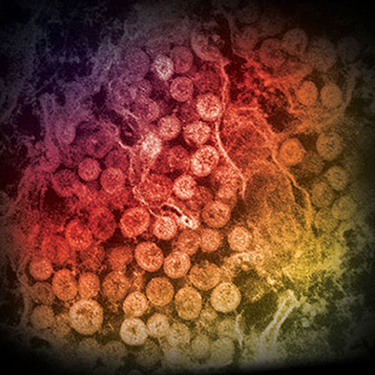
SHL’s Electronic Health Record team are (from left) Jim Sartain, Brian Cook, Dari Shirazi and Alankar Kampoowale. http://tiny.cc/Jan_1Central Lee High School senior Mallory Wills (foreground) works with Marinea Mehrhoff in the Rad Chem laboratory as part of the Student Mentorship Program. http://tiny.cc/Feb_2Laboratorians practice how to identify potential agents of bioterrorism as part of a preparedness workshop hosted by the Hygienic Laboratory. http://tiny.cc/Mar_3 The CDC selected the Hygienic Laboratory and four other state public health laboratories to evaluate a new instrument to use in their influenza real-time PCR clinical test. http://tiny.cc/Apr_4A hydrological buoy floats on West Lake Okoboji. The buoy will collect and relay environmental information about the lake’s water quality every 10 minutes until late fall. http://tiny.cc/May_Robyn Miessler-Kubanek stands by the Hygienic Laboratory’s display in the University of Iowa Mobile Museum. http://tiny.cc/June_06Medical lab tech Kizer Friedley works in Black Hawk County Health Department. Wade Aldous, Disease Control Division director, is CLIA director for the county and the Hygienic Laboratory. http://tiny.cc/July_07 Iowa Department of Public Health announced on Aug. 7 that it had joined with a county health department to investigate an outbreak of influenza A (H3) in a long-term care facility. http://tiny.cc/Aug_08In September, a grandfather contacted the Missouri Department of Agriculture to report an aerial application of a pesticide over a K-12 rural public school. http://tiny.cc/Sept_09Tom and Susan Roeder feed their twin babies with milk from the Mother’s Milk Bank. http://tiny.cc/Oct_10A test developed by the Hygienic Laboratory can detect very low levels of insecticides that have been linked to the decline in bee populations. http://tiny.cc/Nov_11An electron micrograph of a thin section of MERS-CoV, shows the spherical particles within the cytoplasm of an infected cell. Photo courtesy of Cynthia Goldsmith/Azaibi Tamin. http://tiny.cc/Dec_12